Project Report
| Feb 18, 2020
PREVENTION BETTER THAN CURE
By DR KRISHNA R MURTHY | PROJECT LEADER
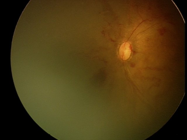
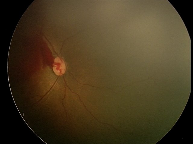
![fundus image of APROP]()
fundus image of APROP
Retinopathy of prematurity (ROP) is related to oxygen-regulated vascular endothelial growth factor and to insulin-like growth factor. The natural course of ROP leads to blindness, causing a social and financial burden on the community. Irreversibly impaired vision may also hinder cognitive and psychomotor development of the affected children.
ROP may be prevented by providing health care for the infant during their stay in the Neonatal Intensive Care Unit (NICU). Oxygen-therapy may be potentially toxic to several organs and tissues, including the still immature retina. Preterm infants are more prone to the effects of oxygen toxicity, since they were used to low oxygen tensions during intrauterine life. After premature birth, there is a dramatic increase in oxygen concentration, which may lead to sustained hyperoxia that may overproduce vascular endothelial growth factor (VEGF). High levels of VEGF stimulate neovascularization of the retina, which in severe cases may result in retinal fibrosis and retinal detachment
Screening programs to detect ROP, including systematic fundus examinations at NICU in infants at risk for ROP, provide the best possibility of diagnosing the disease in order to establish an appropriate treatment prior to progression to more advanced stages and blindness.
- A 3 weeks male baby born in 36 weeks of gestation weighing 800 gms was seen on 10.01.2020 by Dr.Krishna R Murthy. Eye fundus photos of the baby showed aggressive posterior Retinopathy of Prematurity with fovea not yet vascularized (fovea is a tiny pit located in the macula of the retina that provides the clearest vision of all and is aligned with the central axis of the lens). The baby was advised of antivegf injection to both eyes at the earliest. The baby was administered injection on 14.01.2020. As on 14.02.2020; vascularisation still in zone 1 and has been advised for close observation.
- A 6 weeks female baby born in 28 weeks of gestation weighing 1120 gms was seen on 05.12.2019 by Dr.Krishna R Murthy. Eye fundus photos of the baby showed presence of ROP in both eyes in stage 2 in zone 2 in both eyes. The baby was advised of laser intervention to both eyes. The baby underwent laser treatment on 09.12.2019. As on 03.02.2020 the baby is doing well; ROP has regressed in both eyes
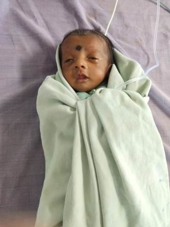
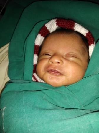
![baby treated for APROP]()
baby treated for APROP
![baby treated with laser therapy]()
baby treated with laser therapy
![Share on Twitter]()
![Share on Facebook]()
Nov 20, 2019
vision therapy
By DR KRISHNA R MURTHY | PROJECT LEADER
![treated with laser for pre retinal haemorrhage]()
treated with laser for pre retinal haemorrhage
The eye works as a camera. The front of the eye has the lens, which focuses on an image, and the pupil, works like a camera shutter to control how much light enters the eye. At the back of the eye is the retina: Like film in the camera, this layer of nerve tissue is necessary to record the information that’s coming in and allow the brain to “develop” it into an image. When babies are born early, the blood vessels that feed the retina usually haven’t finished growing. In ROP, blood vessels swell and overgrow in the light-sensitive layer of nerves in the retina at the back of the eye. These vessels actually stop growing for a time, and then begin growing abnormally and randomly. The new vessels are fragile and can leak, leaving the retina scarred. In the worst-case scenario, the retina detaches (tears away from the back wall of the eye) and puts the baby at high risk of becoming blind.
- A 7 weeks male baby born in 27 weeks of gestation weighing 960 gms was seen on 16.10.2019 by Dr.Krishna R Murthy. Eye fundus photos of the baby showed absence of ROP. The baby was reviewed on 30.10.2019 eye fundus pictures of the baby showed presence of ROP in both eyes in stage 2 in zone 2 (The zone indicates where the disease is located & Zone 2 covers the middle of the retina) with plus disease. The baby was advised of laser intervention to both eyes (Laser therapy burns away the area around the edge of the retina, which has no normal blood vessels). The baby underwent laser treatment on 07.11.2019. As on 20.11.2019 the baby is doing well; ROP has regressed.
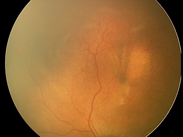
- An 8 weeks male baby born in 28 weeks of gestation weighing 1600 gms was seen on 04.11.2019 by Dr.Krishna R Murthy. Eye fundus photos of the baby showed presence of ROP in both eyes in stage 2 in zone 2 in right eye and in left eye ROP in stage 3 with pre retinal haemorrhage (Neovascularization extends from the ridge into the vitreous). The baby was advised of laser intervention to both eyes. The baby underwent laser treatment on 04.11.2019. As on 20.11.2019 the baby is doing well; ROP has regressed; left eye vitreous haemorrhage is resolving.
![fundus image of pre retinal haemorrhage]()
fundus image of pre retinal haemorrhage
![7 weeks male baby treated with laser]()
7 weeks male baby treated with laser
![Share on Twitter]()
![Share on Facebook]()
Aug 22, 2019
power of sight
By DR KRISHNA R MURTHY | PROJECT LEADER
![2 weeks female baby treated for ROP]()
2 weeks female baby treated for ROP
ROP is a disorder of development of retinal blood vessels in premature babies. Normal retinal vascularization happens centrifugally from optic disc to ora. Vascularization up to nasal ora is completed by 8 months (36 weeks) and temporal ora by 10months (39–41 weeks).The incidence of ROP is increasing in India because of improved neonatal survival rate. The crucial risk factors are – birth weight, gestational age and oxygen therapy. There is need to increase the awareness of the disease to make sure these babies can be treated on time.
1. A 5 weeks female baby born in 27 weeks of gestation weighing 1000 gms was seen on 27.05.2019 by Dr.Krishna R Murthy. Eye fundus photos of the baby showed absence of ROP. The baby was reviewed on 10.06.2019 eye fundus pictures of the baby showed presence of ROP in both eyes in stage 2 in zone 2 with plus disease. The baby was advised of laser intervention to both eyes. Since the stimulus for abnormal vessels comes from the avascular retina therefore ablating the peripheral avascular retina is believed to cause regression of the ROP. The baby underwent laser treatment on 26.06.2019. As on 14.08.2019 the baby is doing well; ROP has regressed.
2. A 2 weeks female baby born in 29 weeks of gestation weighing 1020 gms was seen on 17.07.2019 by Dr.Krishna R Murthy. Eye fundus pictures showed absence of ROP. When the baby was reviewed on 24.07.2019 eye fundus pictures of the baby showed presence of ROP in both eyes in stage 1 in zone 2 with early plus disease. Close observation of the baby was advised. When the baby was reviewed on 31.07.2019; ROP had progressed to stage 2 in zone 2. The baby was advised of laser intervention to both eyes. The baby underwent laser treatment on 03.08.2019. As on 21.08.2019 the baby is doing well; ROP has regressed.
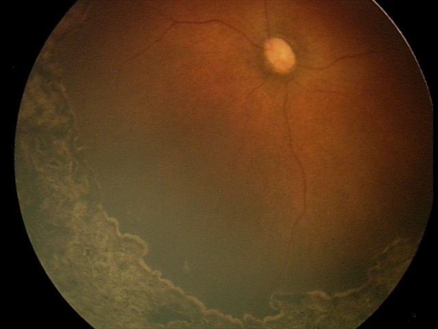
![stage 2 ROP fundus photo]()
stage 2 ROP fundus photo
![fundus photo post ROP laser photo]()
fundus photo post ROP laser photo
![Share on Twitter]()
![Share on Facebook]()