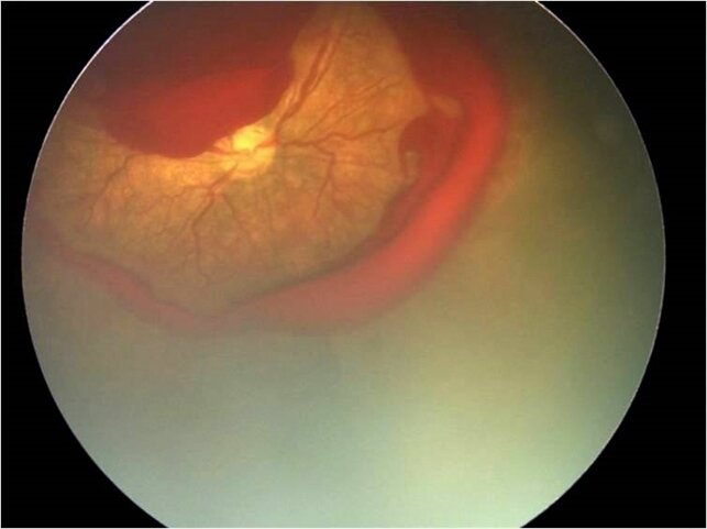
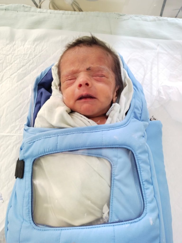
Retinopathy of prematurity (ROP) is abnormal blood vessel development in the retina of the eye. It occurs in infants that are born too early (premature). The blood vessels of the retina (in the back of the eye) begin to develop about 3 months into pregnancy. In most cases, they are fully developed at the time of normal birth. The eyes may not develop properly if a baby is born very early. The vessels may stop growing or grow abnormally from the retina into the back of the eye. Because the vessels are fragile, they can leak and cause bleeding in the eye.
The baby underwent laser treatment on 06.11.2021; as on 14.01.2022 the baby is doing well and ROP has regressed.
The baby underwent laser treatment to right eye and antivegf injection to left eye on 20.11.2021. As on 28.01.2022; ROP in right eye has regressed and in left eye vascularization still present in zone 2 and has been advised to review after 1 week.
Almost all infants with ROP have a gestational age of 31 weeks or less (regardless of birth weight) or a birth weight of 1250 g or less; these indications are generally used to decide whether a baby should be screened for ROP. Any premature baby with severe illness in perinatal period (Respiratory distress syndrome, sepsis, blood transfusion, Intra ventricular haemorrhage, etc.) may also be offered ROP screening.
Retinal examination with scleral depression is generally recommended for patients born before 30–32 weeks gestation, or 4–6 weeks of life, which ever is later. It is then repeated every 1–3 weeks until vascularization is complete (or until disease progression mandates treatment).
Following pupillary dilation using eye drops, the retina is examined using a special lighted instrument (an indirect ophthalmoscope). Examination of the retina of a premature infant is performed to determine how far the retinal blood vessels have grown (the zone), and whether or not the vessels are growing flat along the wall of the eye (the stage). The stage of ROP refers to the character of the leading edge of growing retinal blood vessels (at the vascular-avascular border).
1. A 2 weeks male baby (twin 1) born in 30 weeks of gestation weighing 1280 gms was screened for ROP on 10.05.2021. Eye fundus examination of the baby did not show presence of ROP but incomplete retinal vascular development and areas of peripheral avascular retina was noted and baby was advised for close monitoring. When the baby was reviewed on 24.05.2021 eye fundus examination showed presence of ROP in stage 2 in zone 2 in both eyes with plus disease. The baby was advised for laser treatment to both eyes.
The baby underwent laser treatment on 29.05.2021. As on 07.06.2021 baby is doing well and ROP has refressed.
2. A 3 weeks female baby (twin 1) born in 29 weeks of gestation weighing 1000 gms was screened for ROP on 26.04.2021. Eye fundus examination of the baby showed presence of ROP in stage 2 in zone 2 (Zone 2 covers the middle of the retina) in both eyes with plus disease. The baby was advised for laser treatment to both eyes.
The baby underwent laser treatment on 04.05.2021. As on 17.05.2021 baby is doing well & ROP has regressed.
Premature babies are the number-one risk group for retinopathy of prematurity. In general, the smaller and more premature the infant, the more likely he or she is to develop ROP, and the more likely to need treatment. The eye functions like camera. At the back of the eye is the retina: Like film in the camera, this layer of nerve tissue is necessary to record the information that’s coming in and allow the brain to “develop” it into an image. In premature babies the blood vessels that feed the retina usually haven’t finished growing. ROP occurs when these vessels actually stop growing for a time, and then begin growing abnormally and randomly
A 15 weeks male baby (twin 1st) born in 29 weeks of gestation weighing 1060 gms was reviewed on 25.12.2020 by Dr.Krishna R Murthy. The baby was administered antivegf injection (Antivegf is the use of medications that block vascular endothelial growth factor) to both eyes in the month of November 2020 for APROP (APROP-Aggressive Posterior ROP). On follow-up eye fundus photos of the baby showed presence of ROP in stage 3 in zone 2 in both eyes with plus disease. The baby was advised for laser treatment to both eyes.
In stage 3 ROP the abnormal blood vessels grow toward the center of the eye instead of following their normal growth pattern along the surface of the retina.
The baby underwent laser treatment on 08.01.2021.
The smaller a baby is at birth, the greater their chance of having retinopathy of prematurity (ROP), an eye disorder that can lead to vision loss. But most babies who are born with it get better over time. A baby's eyes begin to develop around the 16th week of pregnancy. If they're born very early, this process is cut short. The blood vessels in their eyes don't have enough time to develop as they should. Instead, they grow where they're not supposed to. Or they can be so fragile that they bleed or leak. ROP can be mild and may resolve spontaneously, but it may lead to blindness in serious cases.
1. A 10 weeks male baby born in 26 weeks of gestation weighing 750 gms was seen on 02.12.2020 by Dr.Krishna R Murthy. Eye fundus photos of the baby showed presence of ROP in stage 2 in zone 2 in both eyes with plus disease. The baby was advised for laser treatment to both eyes. In stage 2 ROP the demarcation line develops into a ridge, with height and width, between the vascular retina and peripheral retina.
The baby underwent laser treatment on 03.12.2020.
2. A 6 weeks female baby born in 29 weeks of gestation weighing 1800 gms was seen on 16.12.2020 by Dr.Krishna R Murthy. Eye fundus photos of the baby showed presence of ROP in stage 3 in zone 1 in both eyes with plus disease. The baby was advised for laser treatment to both eyes at the earliest. Stage 3 is where new blood vessels start to grow out of the ridge. These blood vessels are very weak and they will cause vision problems if they start to scar. In stage 3 ROP the blood vessels grow and multiply (proliferate) and are visible in the ridge
The baby underwent laser treatment on 17.12.2020.
ROP occurs when abnormal blood vessels grow and spread throughout the retina, the tissue that lines the back of the eye. These abnormal blood vessels are fragile and can leak, scarring the retina and pulling it out of position. This causes a retinal detachment. Retinal detachment is the main cause of visual impairment and blindness in ROP.
1. A 9 weeks male baby born in 28 weeks of gestation weighing 1360 gms was reviewed on 19.09.2020 by Dr.Krishna R Murthy. Eye fundus photos of the baby showed presence of ROP in both eyes in stage 2 in zone 2 in both eyes with early plus disease. The baby was advised for laser treatment to both eyes. The baby underwent laser treatment on 27.09.2020. As on 10.10.2020; ROP has regressed and baby is advised to review after 2 weeks.
2. A 4 weeks male baby born in 28 weeks of gestation weighing 1260 gms was seen on 30.09.2020 by Dr.Krishna R Murthy. Eye fundus photos of the baby showed presence of ROP in both eyes in stage 2 in zone 2 in both eyes with plus disease. The baby was advised for laser treatment to both eyes. The baby underwent laser treatment on 01.10.2020.
Project Reports on GlobalGiving are posted directly to globalgiving.org by Project Leaders as they are completed, generally every 3-4 months. To protect the integrity of these documents, GlobalGiving does not alter them; therefore you may find some language or formatting issues.
If you donate to this project or have donated to this project, you will get an e-mail when this project posts a report. You can also subscribe for reports via e-mail without donating.
We'll only email you new reports and updates about this project.