By DR KRISHNA R MURTHY | PROJECT LEADER
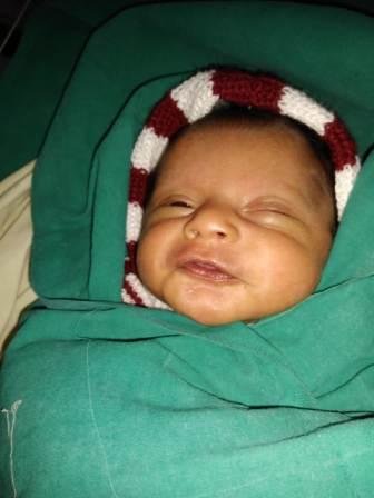
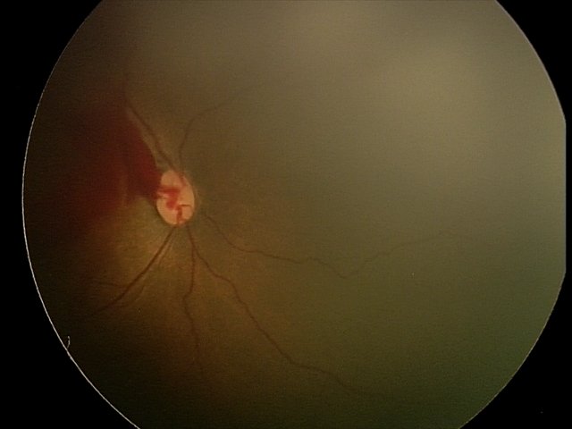
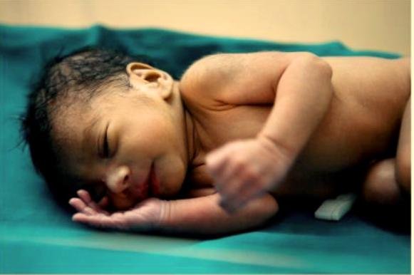
The eye works as a camera. The front of the eye has the lens, which focuses on an image, and the pupil, works like a camera shutter to control how much light enters the eye. At the back of the eye is the retina: Like film in the camera, this layer of nerve tissue is necessary to record the information that’s coming in and allow the brain to “develop” it into an image. When babies are born early, the blood vessels that feed the retina usually haven’t finished growing. In ROP, blood vessels swell and overgrow in the light-sensitive layer of nerves in the retina at the back of the eye. These vessels actually stop growing for a time, and then begin growing abnormally and randomly. The new vessels are fragile and can leak, leaving the retina scarred. In the worst-case scenario, the retina detaches (tears away from the back wall of the eye) and puts the baby at high risk of becoming blind.
Project reports on GlobalGiving are posted directly to globalgiving.org by Project Leaders as they are completed, generally every 3-4 months. To protect the integrity of these documents, GlobalGiving does not alter them; therefore you may find some language or formatting issues.
If you donate to this project or have donated to this project, you can receive an email when this project posts a report. You can also subscribe for reports without donating.