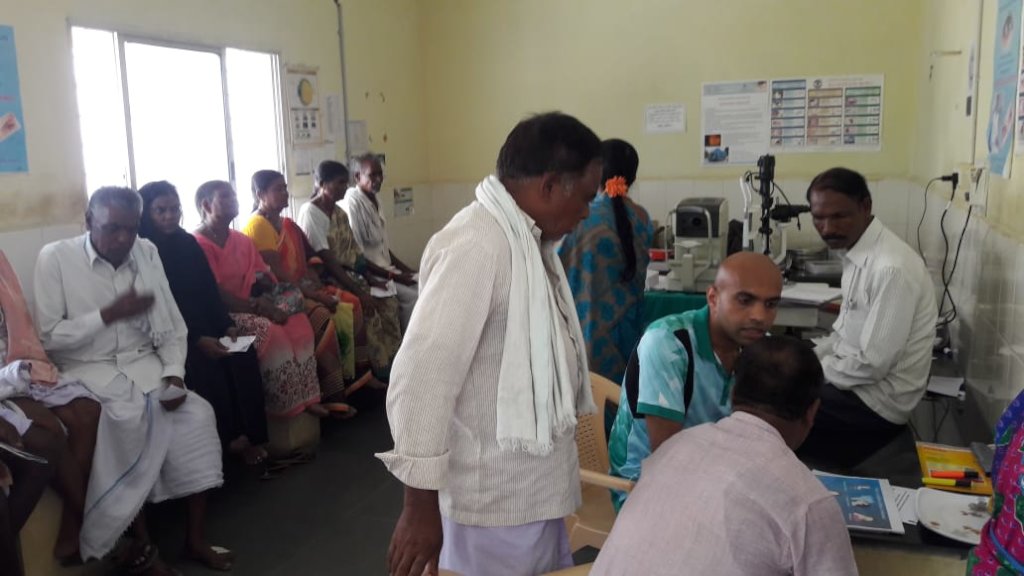
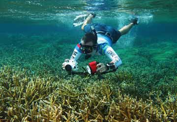
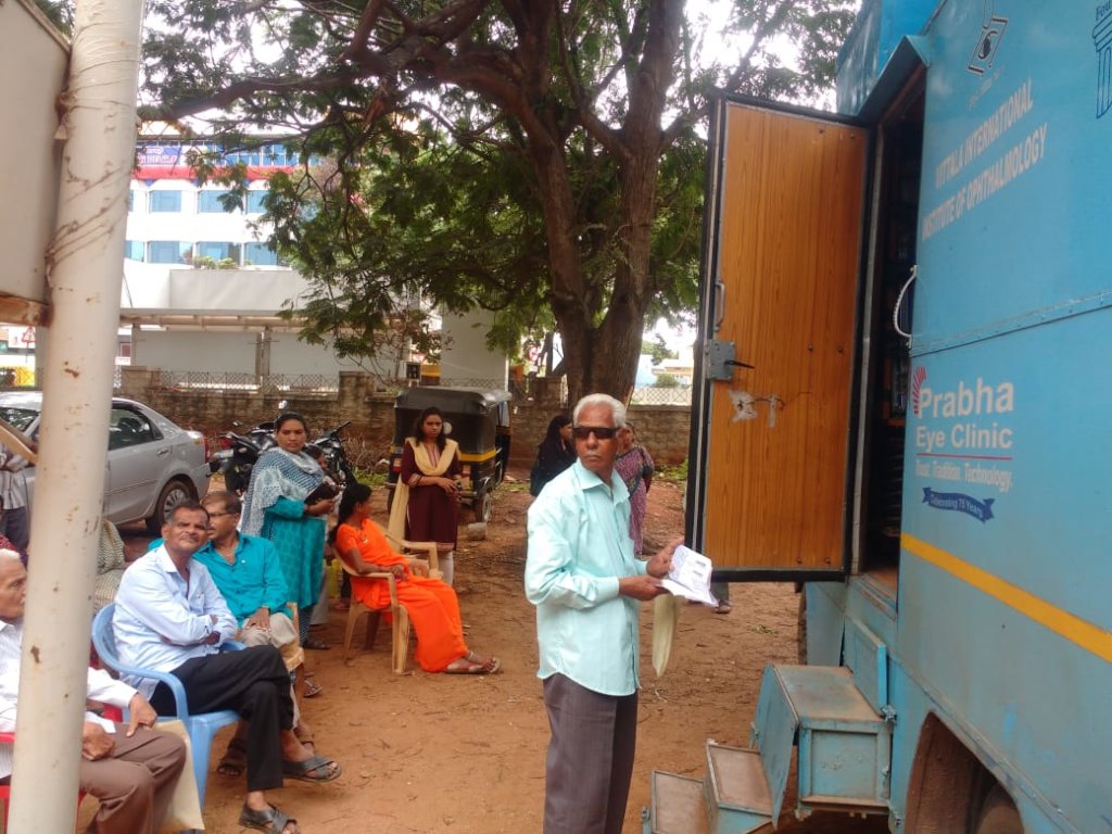
A 58 year old female was referred to us from one of our eye screening activity done at vision centre. She was was seen at our hospital on 08.09.2021 by Dr.Bhargavi Murali, Surgeon and consultant – Vitreo-Retinal Services. On examination she complained of diminution of vision in right eye since many days. She is a known diabetic and hypertensive on treatment for the same. She also has facial nerve palsy and malignant Otitis externa. She was admitted for the management of the same
(Otitis externa is a common ear infection also known as swimmer’s ear. It develops in the ear canal leading to the eardrum. In some cases, otitis externa can spread to surrounding tissue, including the bones of the jaw and face. This infection is known as malignant otitis externa. Malignant otitis externa is an aggressive infection rather than a malignancy, or cancer. An alternative name for malignant otitis externa is necrotizing external otitis. If it’s not treated malignant otitis externa can be life-threatening).
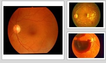
Her visual acuity in right eye was counting fingers at ½ mtr distance. Fundus examination showed advanced with tractional retinal detachment + vitreous haemorrhage. She was advised to undergo RIGHT EYE INTRAVITREAL RAZUMAB INJECTION FOLLOWED BY PARSPLANA VITRECTOMY + MEMBRANE PEELING + ENDOLASER + FLUID GAS EXCHANGE + SILICONE OIL INJECTION UNDER LOCAL ANAESTHESIA.
Surgical Management:
After undergoing all preliminary investigations (physician fitness) She was posted for surgery on 17.09.2021 @ 03:30 pm. The operating team consisted of Dr.Krishna R Murthy & Dr.Bhargavi Murali – Operating surgeons, Dr.Manasi Mehrotra – Assistant Doctor, Mr.Muthuraju - Surgery Assistant, Dr. Raghavendra – Anesthetist, and OT Assistants- Mr.Ravikumar & Mr.Suresh.
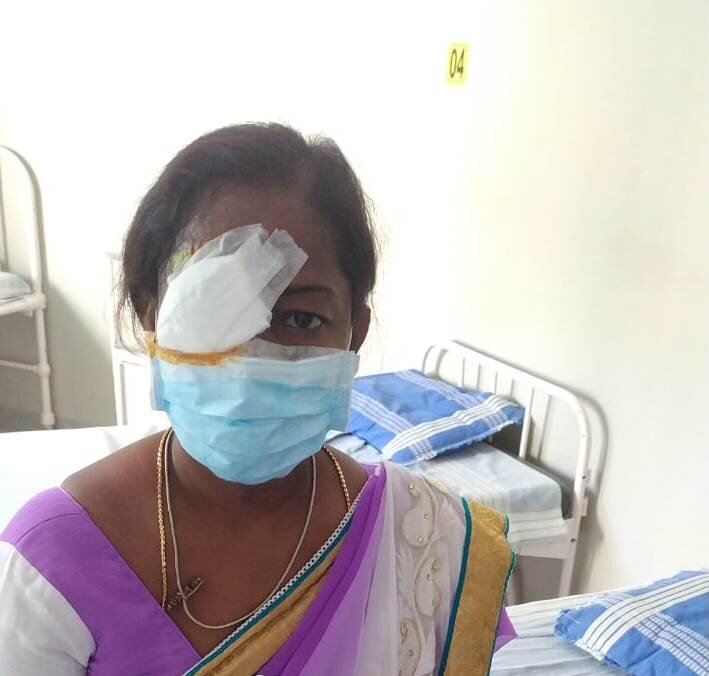
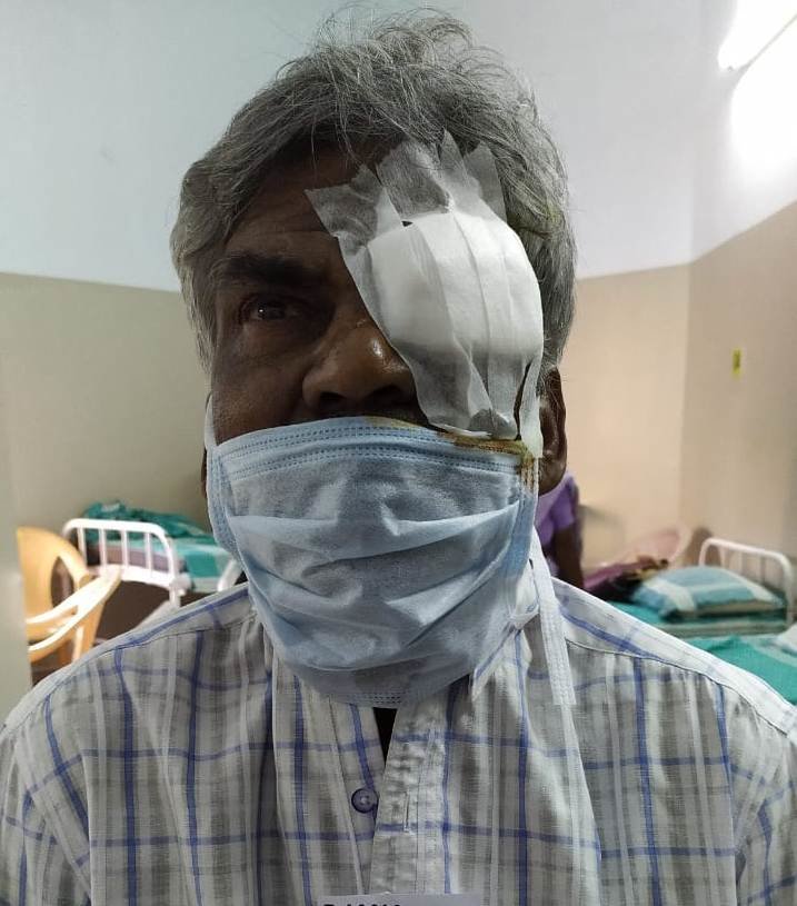
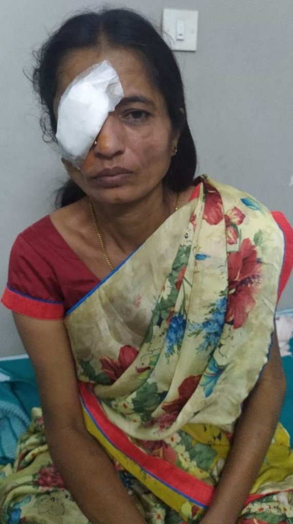
The surgery lasted for 2 hrs 30 min. She was discharged on 18.09.2021 with an advice to follow all medication and to maintain strict prone position for 13-14 hrs a day for 3 weeks and to review after 1 week. As on 08.10.2021, she is doing well; retina is ON.
She is vey happy by her vision status as she is able to do her daily activities on her own, which has helped her gain more confidence for herself.
A 55 yr old gentleman vide MRD # 119612 was seen at our hospital in the month of February 2021. On examination he complained of diminution of vision in both eyes since many years. He is a known hypertensive since 20 yrs on treatment. He had taken retinal laser treatment elsewhere and lost follow-up due to covid 19. His visual acuity in both eyes was presence of perception of light in both eyes. Anterior segment examination showed presence of cataract in both eyes. Fundus examination showed vitreous haemorrhage with retinal detachment in both eyes. He was advised to undergo cataract surgery in left eye followed by surgical intervention for retinal detachment.
He underwent left eye cataract surgery on 09.02.2021. when he was reviewed on 26.02.2021; eye was quiet and cornea was clear and IOL was in place. He was advised to undergo LEFT EYE VITRECTOMY + ENDOLASER + FLUID GAS EXCHANGE + MEMBRANE PEELING + SILICONE OIL INJECTION UNDER LOCAL ANAESTHESIA.
Surgical Management:
After undergoing all preliminary investigations (physical fitness) he was posted for surgery on 02.03.2021 @ 02:00 pm. The operating team consisted of Dr.Vinaya Kumar K – Operating surgeon, Dr.Manasi – Assistant Doctor, Mr.Umesh - Surgery Assistant, Dr. Naveen – Anesthetist, and OT Assistants- Mr.Mahadevaswamy & Mr.Samuel Dennis.
The surgery lasted for 2 hrs. He was discharged on 03.03.2021 with an advice to maintain strict prone position for 13-14 hrs a day for 3 weeks and review after 1 week. As on 30.03.2021; he is doing well and retina is ON and has been advised to review after 2 weeks. His visual acuity in left is counting fingers at 2 mtr distance.
He is basically a flower vendor. He supports his sister in this work. He is satisfied with the surgery outcome and is happy that he is able to extend his helping hands to his sister who is a caretaker.
The retina is a complex transparent tissue consisting of several layers, only one of which contains light-sensitive photoreceptor cells. Light must pass through the overlying layers to reach the photoreceptor cells, which are of two types, rods and cones. Rods in humans provide night vision and aid in visual orientation. Cones are more prominent in humans and provide detailed vision (as for reading) and colour perception.
Under our project complex retinal diabetic surgeries we have operated VITRECTOMY SURGERY on a 46 yr old Female.
She has been under our care and management since 2007 for management of CMV retinitis (Cytomegalovirus retinitis (CMV retinitis) is a serious viral eye infection of the retina. The retina is the light-sensing nerve layer that lines the back of the eye. It is most often found in people with weakened immune systems. This can lead to a loss of peripheral (side) vision. Sometimes the symptoms begin with a blind spot in the center of vision and can lead to a loss of central vision. The symptoms usually happens first in one eye but often progress to the other eye. Without treatment or improvement in the immune system, CMV retinitis destroys the retina and damages the optic nerve. This results in blindness).
When she was reviewed in Feb 2021 in right eye showed healed CMV retinitis with nasally detaching retina. She was advised for surgical intervention of RIGHT EYE VITRECTOMY + MEMBRANE PEELING + ENDOLASER + FLUID GAS EXCHANGE + ENDOLASER + C3F8 GAS UNDER LOCAL ANAESTHESIA.
Surgical Management:
After undergoing all preliminary investigations (physical fitness) she was posted for surgery on 05.03.2021 @ 02:15pm. The operating team consisted of Dr.Krishna R Murthy & Dr.Bhargavi Murali – Operating surgeons, Dr.Raheela Afsan – Assistant Doctor, Mr.Anil Kumar - Surgery Assistant, Dr. Raghavendra – Anesthetist, and OT Assistants- Mr.Ravikumar & Mr.Mahadevaswamy.
The surgery lasted for 2 hrs 30 min. She was discharged on 06.03.2021 with an advice to follow all medications and to maintain strict prone position for 13-14 hrs a day for 3 weeks and review after 1 week. As on 26.03.2021; she is doing well and retina is ON and is suggested to review after 3 weeks.
She works in a private firm as a helper staff and supports her family of 3 members. Husband deceased years back and the responsibility of children lies on her who are still pursuing their graduation studies. The above support has helped her to great extent.
The retina is the part of the eye that receives the light and converts it into chemical energy. The chemical energy activates nerves that conduct the messages out of the eye into the higher regions of the brain. The retina is a complex nervous structure.
Under our project complex retinal diabetic surgeries we have operated VITRECTOMY SURGERY on a 47 yr old gentleman.
He was seen at our hospital in the month of June 2020 by Dr.Krishna R Murthy. On examination he complained of diminution of vision in left eye since 8 months & gave of history of trauma to his right eye with stone 35 yrs back. Fundus examination in right eye showed total glaucomatous disc and in left eye posterior retinal detachment. (A retina detaches when the retina peels away from its underlying layer of support tissue at the back of the eye). He was advised to undergo LEFT EYE VITRECTOMY + ENDOLASER + FLUID GAS EXCHANGE + SILICONE OIL INJECTION UNDER LOCAL ANAESTHESIA.
Surgical Management:
After undergoing all preliminary investigations (physical fitness) he was posted for surgery on 20.10.2020 @ 02:15pm. The operating team consisted of Dr.Krishna R Murthy & Dr.Bhargavi Murali – Operating surgeons, Dr.Snehapriya – Assistant Doctor, Mr.Umesh - Surgery Assistant, Dr. Naveen – Anesthetist, and OT Assistants- Mr.Suresh & Mr.Muthuraj,
The surgery lasted for 2 hrs 30 min. He was discharged on 21.10.2020 with an advice to follow all medications and to maintain strict prone position for 13-14 hrs a day for 3 weeks and review after 1 week. As on 10.11.2020; he is doing well and retina is ON and was suggested to review after 3 weeks for glasses suggestion.
By profession he works in a hotel industry as a helper and supports his family. The above helping hand has imbibed confidence in him to get involved in some job to support his family.
Background:
A 53 year old gentleman was reviewed at our hospital on 17.01.2020 by Dr.Bhargavi Murali. He had undergone both eyes cataract surgery in 2015. On examination he complained of diminution of vision in right eye since 4 days. His visual acuity in right eye was counting fingers at 1 mtr distance and in left eye 6/6p. Fundus examination in right eye showed inferior retinal detachment with macula off (It is the status of the macula that defines the urgency with which surgical repair should be undertaken, Macular detachment is a detachment involving the fovea with any resulting loss of central Snellen visual acuity).
He was advised to undergo RIGHT EYE VITRECTOMY + ENDOLASER + FLUID GAS EXCHANGE + ENDOLASER UNDER + C3F8 GAS UNDER LOCAL ANAESTHESIA.
Surgical Management:
After undergoing all preliminary investigations (physical fitness) he was posted for surgery on 21.01.2020 @ 02:30pm. The operating team consisted of Dr.Krishna R Murthy – Operating surgeon, Dr.Snehapriya – Assistant Doctor, Mr.Anilkumar - Surgery Assistant, Dr. Naveen – Anesthetist, and OT Assistants- Mr.Suresh & Mr.Ravikumar,
The surgery lasted for 1 hr 45 min. He was discharged on 22.01.2020 with an advice to follow all medications and to maintain strict prone position for 13-14 hrs a day for 3 weeks and review after 1 week. As on 21.02.2020; he is doing well and retina is ON and was suggested to review after 3 weeks.
By profession he works as a school van driver and supports his family. His profession requires him to have correct eyesight for safe driving. Post surgery his visual acuity in right eye is 6/12. Timely intervention has helped him to continue his profession and support his family
Project reports on GlobalGiving are posted directly to globalgiving.org by Project Leaders as they are completed, generally every 3-4 months. To protect the integrity of these documents, GlobalGiving does not alter them; therefore you may find some language or formatting issues.
If you donate to this project or have donated to this project, you can recieve an email when this project posts a report. You can also subscribe for reports without donating.