By Dr Krishna R Murthy | Project Leader
Background
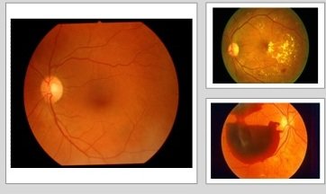
A 31 yr old male gentleman was seen at our hospital on 15.07.2019 by Dr.Bhagya. He was treated elsewhere for infective retinitis with topical and systemic medications but with no significant improvement. His left eye vision was progressively deteriorating and he presented with visual acuity of counting fingers close to face in left eye and in right eye was 6/18. Anterior segment examination showed fibrin membrane in pupillary area. Fundus examination showed dense vitritis in right eye and with no view in left eye. He was advised for some special blood investigation and was maintained on topical steroid management. Even with topical steroid management his visual acuity in left eye still deteriorated to hand movements close to face. He was advised for RIGHT EYE VITRECTOMY + ANTERIOR CHAMBER VITREOUS TAP FOR FUNGAL, HISTOPATHOLOGY AND KOH GREAM STAIN INVESTIFATIONS + INTRAVIT ANTIBIOTIC AND ANTIFUNGAL INJECTIONS UNDER LOCAL ANAESTHESIA.
Surgical Management:
After undergoing all preliminary investigations (physical fitness) he was posted for surgery on 26.07.2019 @ 02:30pm. The operating team consisted of Dr.Krishna R Murthy & Dr.Bhargavi Murali – Operating surgeons, Dr.Anusha – Assistant Doctor, Mr.Suresh - Surgery Assistant, Dr. Raghavendra – Anesthetist, and OT Assistants- Mr.Muthuraju & Mr.Anilkumar. The anterior chamber tap was sent to outsourced lab for the required investigations.The anterior chamber tap came positive for pan fungal genome and he was treated with multiple doses of antifungal intravitreal injections. He was also sent for systemic evaluation to rule out systemic vasculities.
The surgery lasted for 1 hr 15 min. He was discharged on 27.07.2019 with an advice to follow all medications and to review after 1 week. He was on regular and close follow-up post surgery. His visual acuity started improving gradually and as on 25.09.2019; he is doing fine and his visual acuity in right left eye is 6/6.
Uveitis is the inflammation of the uvea, the pigmented layer that lies between the inner retina and the outer fibrous layer composed of the sclera and cornea. Iridocyclitis is an inflammation of the iris (the colored part of the eye) and of the ciliary body (muscles and tissue involved in focusing the eye.
He and his family are dependent on the produce from the agricultural land. He owned around 1 acre of agricultural land on which he used to reap Jowar and millet. Since past 3 years he had been running around to eye hospitals for his eye problem. The expenses of the investigations forced him to sell some portion of his agricultural land. He was very much depressed due to uncertain treatment and management done till now and due to his deteriorating vision he was unable to earn and support the family. Relatives and friends pitched in with some financial support and they guided him to our hospital.
He is very happy with the improvement in his vision and he is able to carry on his regular work activities. He thanked the entire team for the outcome.
Project reports on GlobalGiving are posted directly to globalgiving.org by Project Leaders as they are completed, generally every 3-4 months. To protect the integrity of these documents, GlobalGiving does not alter them; therefore you may find some language or formatting issues.
If you donate to this project or have donated to this project, you can receive an email when this project posts a report. You can also subscribe for reports without donating.