By DR.KRISHNA R MURTHY | PROJECT LEADER
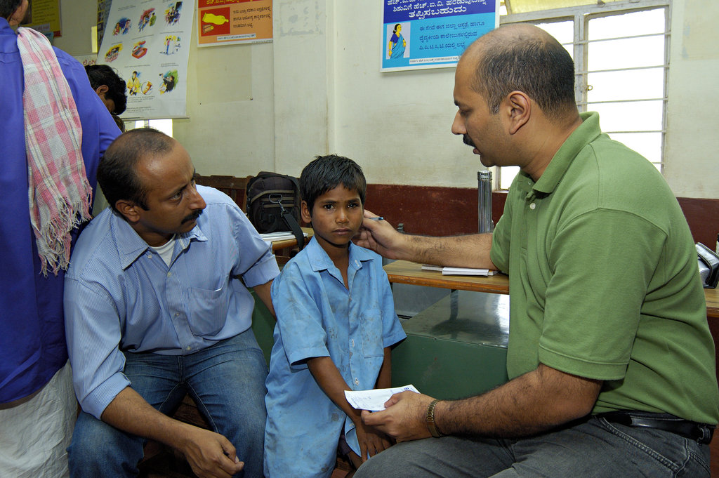
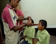
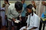
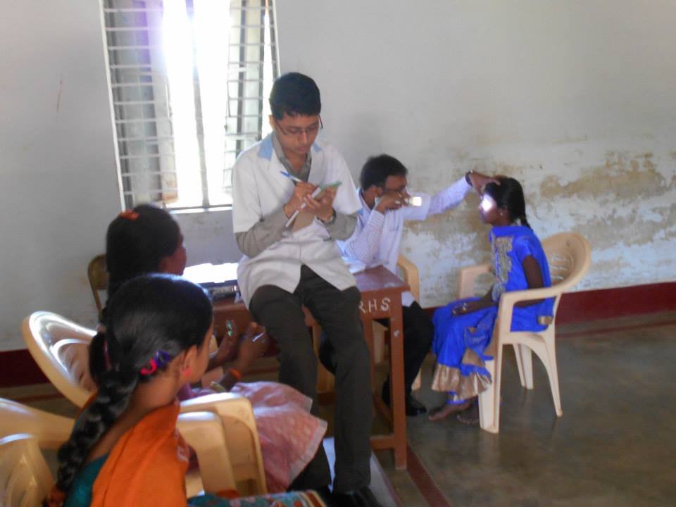
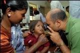
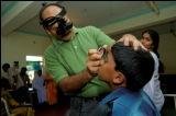
In accordance to our driving spirit to fulfill the dream of our founder, we are continuously striving to spread awareness and educate regarding childhood vision problems and its side effects in children. A child needs many abilities to succeed in school. Good vision is a key. 80% of the learning a child does occurs through his or her eyes. Reading, writing, and using computers are among the visual tasks students perform daily. Healthy eyes and vision are a critical part of children development to excel in their curricular activities. To make this effort successful Parents and teachers are educated the need to be alert for symptoms that may indicate a child has a vision problem. Under this effort we have successful treated the below children for the following eye problems.
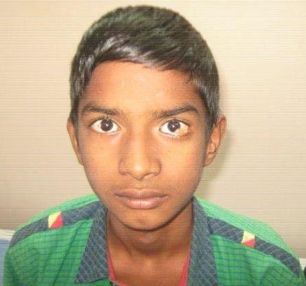
1. Paediatric anterior uveitis - Uveitis is a condition causing inflammation of the middle eye or uvea) + Juvenile Idiopathic Arthritis (Juvenile idiopathic arthritis (JIA) can be defined as a group of idiopathic arthritides, which occurs before the age of 16 years and persists for at least 6 weeks. It is the most common cause of uveitis in children and also major cause of visual impairment in children). Bilateral lensectomy was done to treat the child.
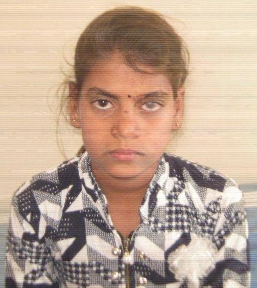
2. Limbal dermoid- They are usually superficial lesions. They appear most frequently at the inferior temporal quadrant of the corneal limbus. Limbal dermoids are benign congenital tumors that contain choristomatous tissue (tissue not found normally at that site). MRI scan of brain and orbit will be done to see the extention and involvement of eye muscles. Limbal dermoid excision with histopathology examination was done to treat the child.
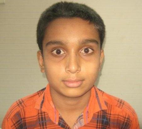
3. Herpes zoster & keratitis - Herpes zoster ophthalmicus is reactivation of a varicella-zoster virus infection (shingles) involving the eye. Symptoms and signs, which may be intense, include dermatomal forehead rash and painful inflammation of all the tissues of the anterior and, rarely, posterior structures of the eye, Shingles in the eye could lead to vision loss). Penetrating Keratoplasty was the remedial surgical measure taken to restore the vision for the child.
4. Keratoconus - It is a degenerative disorder of the eye in which structural changes within the cornea cause it to thin and change to a more conical shape than the more normal gradual curve. Keratoconus can cause substantial distortion of vision, with multiple images, streaking and sensitivity to light. It is typically diagnosed in children in the age group of 10-18 yrs. C3R (COLLAGEN CROSS-LINKING) SURGERY was the remedial measure taken to stabilize the cornea and vision for the child.
Project reports on GlobalGiving are posted directly to globalgiving.org by Project Leaders as they are completed, generally every 3-4 months. To protect the integrity of these documents, GlobalGiving does not alter them; therefore you may find some language or formatting issues.
If you donate to this project or have donated to this project, you can receive an email when this project posts a report. You can also subscribe for reports without donating.