By DR.KRISHNA R MURTHY | PROJECT LEADER
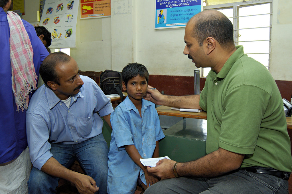
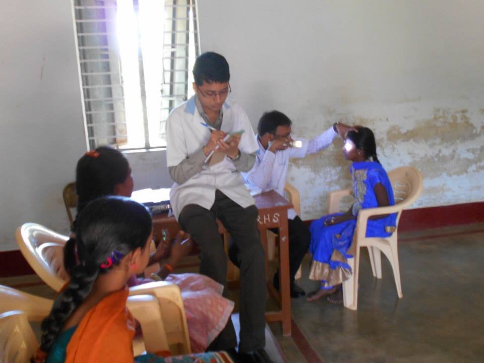
Healthy eyes and vision are a critical part of child development. Signs that a child may have vision problems include constant eye rubbing, extreme light sensitivity, poor focusing, poor visual tracking (following an object) abnormal alignment or movement of the eyes (after 6 months of age) chronic redness of the eyes, chronic tearing of the eyes, a white pupil instead of black. Regular eye exams can detect and correct this and other vision problems.
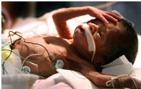
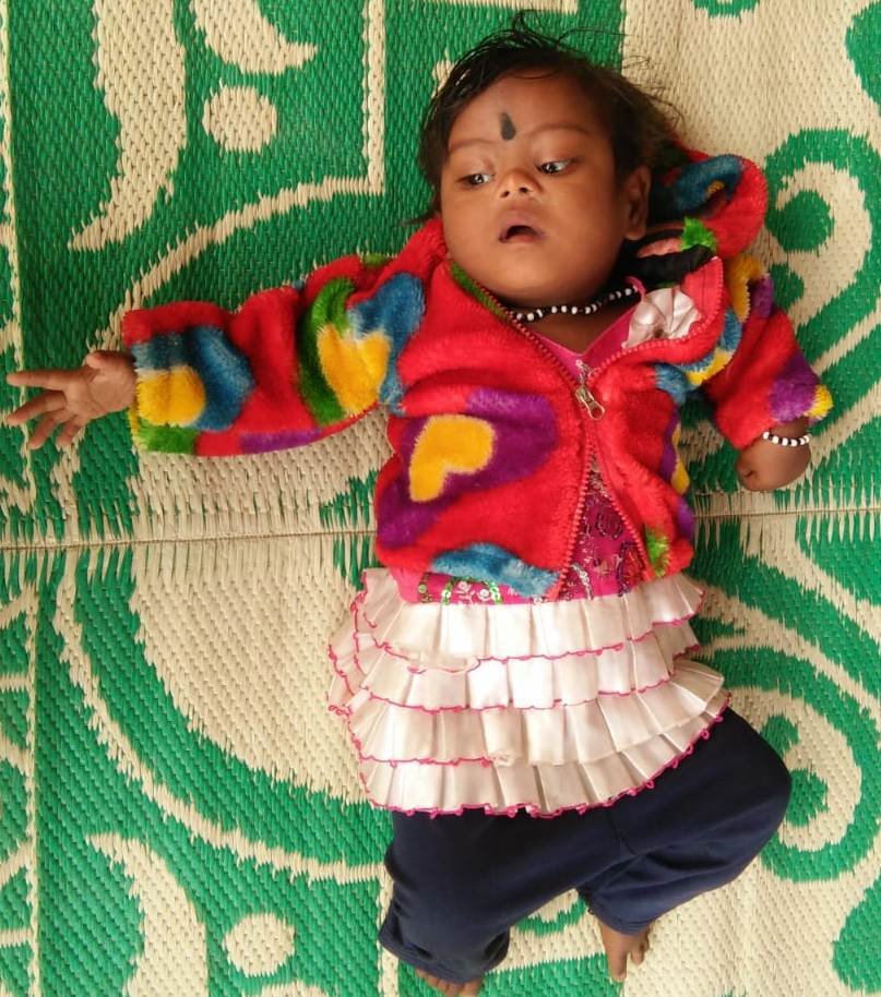
CONGENITAL CATARACT: cataract present at birth.
A 7 months old female child was seen by us in the month of August 2018; the baby was under treatment for Down’s syndrome and congenital heart disease. Down syndrome is a chromosomal condition that is associated with intellectual disability, a characteristic facial appearance, and weak muscle tone (hypotonia) in infancy. Children with Down syndrome have an increased risk of developing several medical conditions and have an increased risk of hearing and vision problems. On examination the baby was able to follow light well in all directions. The baby was diagnosed to have calcified cataract with Nystagmus. Nystagmus is a vision condition in which the eyes make repetitive, uncontrolled movements. The child was advised for BOTH EYES CATARACT EXTRACTION + ANTERIOR VITRECTOMY + PARSPLANA CAPSULOTOMY UNDER GENERAL ANAESTHESIA. The child underwent the surgery on 21.12.2018.
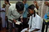
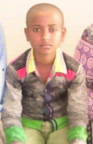
CORNEAL SCAR: Injury to cornea - The cornea is the eye’s outermost layer. It is the clear, domeshaped surface that covers the front of the eye. It plays an important role in focusing our vision. The cornea and lens of the eye are built to focus light on the retina, which is the light-sensitive tissue at the back of the eye. When light strikes the cornea, it bends—or refracts—the incoming light onto the lens. The lens refocuses that light onto the retina, which starts the translation of light into vision. Minor injuries or scratches, the cornea usually heals on its own. Deeper injuries can cause corneal scarring, resulting in a haze on the cornea that impairs vision.
An 11 yrs old boy was presented to us with the above condition He came to us with diminution of vision in left eye; his visual acuity in left eye was counting fingers at ½ mtr distance. Anterior segment examination showed corneal scar which necessitated surgical intervention of LEFT EYE KERATOPLASTY UNDER GENERAL ANAESTHESIA. The child underwent the surgery on 07.01.2019. As on 07.02.2019; the child is doing well, his visual acuity in left eye is counting fingers at 2 mtr distance.
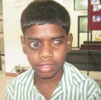
CONGENITAL GLAUCOMA: Congenital glaucoma is a developmental glaucoma that results from the abnormal development of the aqueous drainage structure, characterized by an elevated intra-ocular pressure, enlargement of globe (buphthalmos), corneal edema and optic nerve cupping.
A 14 yr old boy has been under our care and management for congenital glaucoma since 2011. The child has undergone multiple surgical interventions to retain the vision in the only Seeing Eye – right eye. On follow-up on 07.01.2019; the intraocular pressure (IOP) in right eye was 34 mmHg which is a sight threatening condition. The child was advised to undergo RIGHT EYE AADI (AUROLAB AQUEOUS DRAINAGE IMPLANT) UNDER GENERAL ANAESTHESIA. The child underwent the same on 31.01.2019. As on 07.02.2019; IOP is well controlled and his visual acuity is counting fingers at 2.5 mtr.
Project reports on GlobalGiving are posted directly to globalgiving.org by Project Leaders as they are completed, generally every 3-4 months. To protect the integrity of these documents, GlobalGiving does not alter them; therefore you may find some language or formatting issues.
If you donate to this project or have donated to this project, you can receive an email when this project posts a report. You can also subscribe for reports without donating.