By DR KRISHNA R MURTHY | PROJECT LEADER
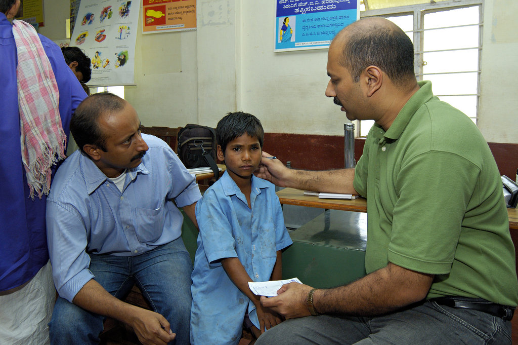
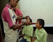
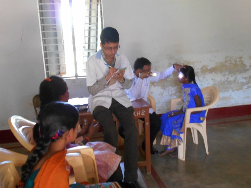
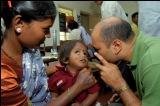
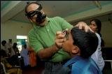
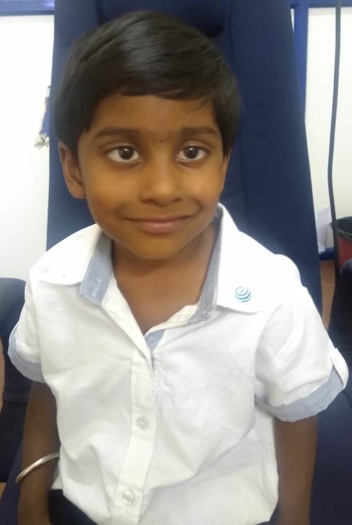
Most of the children in their school going age are unaware of their visual needs or vision related problems. The proverb “an ounce of prevention is worth a pound of cure” is true with children’s eye care. As children progress throughout their education, they face increasing demands on their visual abilities. When certain visual skills have not developed, or are poorly developed, learning is difficult and stressful. Children will typically attempt to do the work, but with a lowered level of comprehension or efficiency. Eye exams by an eye doctor are an important way to identify problems with your child's vision. Problems that are found early have a better chance of being treated successfully
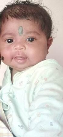
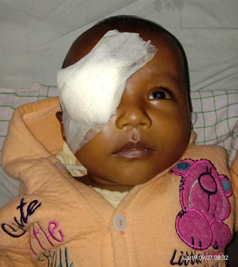
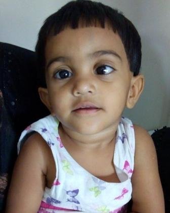
CONGENITAL GLAUCOMA:
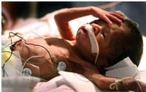
A 7 months old male baby was seen by us on 31.10.2019 in Glaucoma Department. The baby has the systemic condition called sturge weber syndrome with seizure. It is a congenial disorder and a rare neurological condition that affects the development of certain blood vessels causing abnormalities in the brain, skin and eyes from birth. It is characterized by a port-wine birthmark on the child’s face. On examination parents informed of treatment at Indira Gandhi Institute of Child Health care for epilepsy since 1 month. During visual acuity examination the child was able to follow and fix light in both eyes. Anterior segment examination showed megalo cornea in both eyes. An intraocular pressure in right eye was 24 mmHg and in left eye was 28 mmHg. The child was advised to undergo BOTH EYES EXAMINATION UNDER ANAESTHESIA + TRABECULECOTMY + TRABECULOTOMY UNDER GENERAL ANAESTHESIA. The child was advised to start perioperatively oral propranalol 1 week prior to surgery (to treat high blood pressure and circulatory conditions after pediatrician opinion)
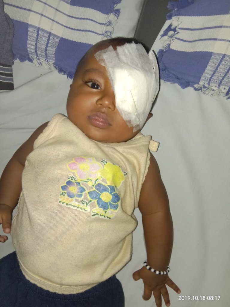
After undergoing all preliminary investigations (paediatrician fitness) the baby was posted for surgery on 30.12.2019. The surgery lasted for 1 hr 45min and was uneventful.
As on 21.01.2020; the baby is doing well and examination under anaesthesia has been advised.
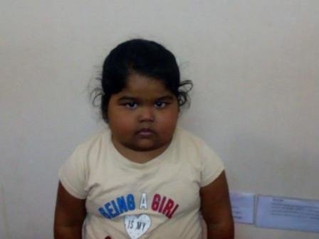
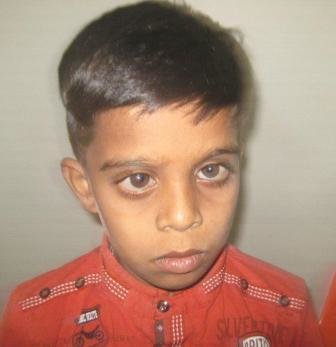
DEVELOPMENTAL CATARACT
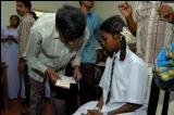
A 7 year old female child was seen at our hospital in the month of September 2019. On examination parents gave a history of undergoing cataract surgery to left eye 2 yrs back elsewhere. The child complained of diminution of vision in right eye. Her visual acuity in right eye was counting fingers at 1 mtr distance and in left eye was 6/60. Anterior segment examination showed presence of cataract with diffuse lenticular opacities. The child is suffering from chronic disease known as nephritic syndrome. (Nephritic syndrome is the name given to a collection of different signs and symptoms that occur as a result of inflammation in the kidneys. This inflammation causes the kidneys to works less effectively. It also causes protein and red blood cells to leak from the blood stream into the urine. There are many conditions that may cause nephritic syndrome and it can occur in people of all ages. Common causes are infections, immune system disorders and inflammation of the blood vessels. The main symptoms are passing less urine than normal, leading to a fluid buildup in the body, and having blood in the urine. People with nephritic syndrome also often develop high blood pressure). The child is under treatment for the above condition. The child was advised to undergo RIGHT EYE CATARACT EXTRACTION + ANTERIOR VITRECTOMY + IOL IMPLANTATION + POSTERIOR CAPSULOTOMY UNDER GENERAL ANAESTHESIA. Post surgery close monitoring on child’s BP fluctuations was advised by the paediatrician and was monitored round the clock.
After undergoing all preliminary investigations (paediatrician fitness) the child was posted for surgery on 05.10.2019
By DR KRISHNA R MURTHY | PROJECT LEADER
By DR.KRISHNA R MURTHY | PROJECT LEADER
Project reports on GlobalGiving are posted directly to globalgiving.org by Project Leaders as they are completed, generally every 3-4 months. To protect the integrity of these documents, GlobalGiving does not alter them; therefore you may find some language or formatting issues.
If you donate to this project or have donated to this project, you can receive an email when this project posts a report. You can also subscribe for reports without donating.